Pathologists would gain new tool to diagnose cancer faster and more accurately, based upon stain-free analysis of tissue
Reading tissue biopsies with a new stain-free method could eventually help pathologists achieve faster and less subjective cancer detection. Should this technology prove viable, it would also displace many of the longstanding tissue preparation methodologies used today in the histopathology laboratory.
Credit a research team from the Beckman Institute at the University of Illinois (UI) Christie Clinic and at the UI campuses in Urbana and Chicago, with developing this new technology.
They call the technique Spatial Light Interference Microscopy (SLIM). According to a story reported by Futurity.org, the technique uses two beams of light.
New Technology Could Help Pathologists Detect Cancer Earlier
In the Proceedings of the National Academy of Sciences, the scientists stated the new technology offers answers to some of the most elusive questions in contemporary biology: how cell growth is regulated and how cell size distributions are maintained. “SLIM can be so valuable for greatly improving the chances of early detection and treatment of cancer,” declared study leader Gabriel Popescu, Ph.D., Quantitative Light Imaging Laboratory, Department of Electrical and Computer Engineering at the Beckman Institute.
The reason for Popescu’s optimism is SLIM’s capabilities using optical interferometry, or interference patterns, to make accurate measurements of waves at the molecular level. This enables the technique to work with great sensitivity.

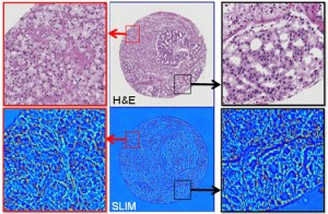
Shown above are multi-modal
images of prostate biopsy slides of a 5+4 Gleason grade, or high grade
tumor. The top row of images are done with standard histological
staining. The bottom row of images are done with the stain-free Spatial
Light Interference Microscopy (SLIM). (Credit: Shamira Sridharan of www.Futurity.org)
According to Mustafa Mir,
a graduate student in Electrical Engineering and a first author of the
project paper, SLIM is capable of measuring mass with a sensitivity of
one femtogram, or one thousandth of the mass of a cubic micron of water.“What that means is that only a small number of molecules arranged in a certain way are enough to give us the optical signal that something is going to happen here,” Popescu explained.
The technology works through a combination of phase-contrast microscopy and holography. It does not need staining or any other special preparation of the tissue to be analyzed. It is also completely non-invasive. This means that scientists can visualize nanoscale structures quantitatively and study ongoing cell function in situ.
“Ideally, we would like to detect cancer at the single-cell level,” Popescu stated. This would enable scientists to find a cell that looks abnormal early on, allowing treatment where the process is still reversible. “We know that the disease starts at the nanoscale, at the molecular level, and we think we have the proper tool to catch these early events,” he added.
With Current Staining Method, Pathologists Disagree 20% of the Time
SLIM offers revolutionary advantages over current staining technology. Its optical maps report morphological properties of tissues and cells that cannot be recovered by common stains, including hematoxylin and eosin, Futurity reported. This provides objective evaluations of structural data such as tumor margins that can be difficult for pathologists to assess with current methodologies.
“A significant advantage over existing methods is that we can measure all types of cells… while maintaining the sensitivity and the quantitative information that we get,” Mir stated in a story by MedGadget.
According to Popescu, use of SLIM technology, along with a fluorescent reporter, could have broader implications in understanding the effects of cancer treatments and other forms of therapy on the fundamental process of cell growth. “By using [the combined technologies], we were also able to differentiate how the cells regulate their growth in different stages of their lifecycle,” he stated.
“We think that the most important advantage of SLIM is that it provides quantitative, objective information,” Popescu observed in Futurity. “Right now, in the clinic, the diagnosis is subjective; it’s a human that does it. There are studies showing that two pathologists agree on a diagnosis only four out of five times.”
Popescu described the ability to actually predict information about the outcome of the patient as the Holy Grail of the research. As an example, he noted SLIM’s potential to help determine the likelihood of recurrence following surgery. “This, right now, is actually a 50/50 guess,” he observed.
According to Popescu, SLIM’s highly automatic procedure, together with the low cost and high-speed associated with the absence of staining, could make “a significant impact in pathology at a global scale.”
Of course, it will take years before tissue analysis solutions incorporating Spatial Light Interference Microscopy (SLIM) are ready for daily use by anatomic pathologists. Yet, it is the pipeline of transformative technologies like that which promise to give Clinical laboratory professionals new capabilities to diagnose disease more accurately and earlier. In turn, this improves the value that laboratory medicine brings to physicians, patients, and payers.
—Pamela Scherer McLeod
Source