Sunday, September 14, 2014

Manali to Leh: A Bicycle Tour

Manali to Leh is a dream route for most cycling enthusiast.The route presents challenges of high altitude, freezing temperatures, uncertain weather, and long stretches of winding roads. Despite all these and more known difficulties when we started to plan cycling on this route, five of us fellow cyclists decided to join, all from Bhuj Bicycling Club. Me, Dr. Devanand Parmar, Shreyas Negi, Mahendra Yadav and Bhavesh Dave committed for the job. We started to prepare about 3 months before the planned date of 22nd August. Our daily training included about 20-30 km of cycle riding in early morning and about 50 km of riding on Sundays. However we later realized that no training can prepare one for the rigours of the Manali Leh route. Anyway, soon this time passed and the day of journey arrived.


Starting from Bhuj, on 19th August

Sunday, December 29, 2013

THE GOD: A pathologist's view


(Disclaimer: Following article is written in one go. It may lose flow, or may become difficult to understand. This article is not copied from anywhere and it is authors own genuine thinking. If you want to publish this article or part of the article somewhere else kindly give proper credit and information to the author.)


Our body is made of cells. Cells are considered as the smallest unit of the body that can survive outside human body if it's needs are fulfilled. To give an example we can keep cells in the donated blood alive for a month, and if we can transfuse these cells to other human, the cells in donated blood will survive. Nowadays we can even culture some of the human cells outside the human body.


The soul (or atma) of a human is said to be the life force. It is immortal and unbreakable. It changes body like body change clothes. Without soul body will be dead. That means without soul all the cells of the body will die.


If we combine above two paragraphs, we can find few controversies. When we remove red blood cells from a blood donor, do we remove part of his soul? If not, how these cells can survive without soul? When to transfuse these red cells to a recipient, do we transfuse cells with part of donor's soul? How can we culture human cells ( in culture we grow human cells, from one to many), if there is no soul or life force in those cells?


Explanation:


Above problem can be explained if we consider that every cell carries it's own life force or it's own soul. Like when we combine all the cells of the body, we make one human. When we combine all the life force of all cells we make soul of human. So soul of human is made by combining souls of all the cells. So when we remove cells from a donor we are removing life force of that cells to, so these cells can survive outside human body.


If one human soul or life force can be made of multiple life forces, we may further theorise that if we combine life forces of all the living organism, we make an ultimate life force, that can be called GOD.


By this theory The God is not a single being, but is made of all the life force this world has. So when majority of do something, to bring change in the world, if becomes The God's decision.


Purnamadah Purnamidam Purnat Purnmudachyate

Purnasya Purnmaday Purnmevmashisyati.


When we remove few red cells from a donor, we are not breaking soul or life force. The life forces of all the living organisms are connected with each other in the form of the God. And despite these every single cell, whether it is a cell from human, or it is a bacteria, is complete in it's self. Everyone is complete like the God.


Wednesday, March 6, 2013

Bio disc party trick, How?

Click here to know
Just click above to check few videos about how the finger lifting party trick demonstrated by Bio disc salesman works.

Sunday, January 6, 2013

GMC guidelines regarding Continuing Medical Education and Credit Hours


1. PREAMBLE

Medical Science is dynamic and it is essential for the doctor to become acquainted with the advances in medicine. This is in essence the concept of Continuing Medical Education (CME). Tremendous advances are taking place in the field of medical sciences, continuously changing the concept, approach to management and the outcome of several diseases. The rapid pace of these advances makes it mandatory for doctors to keep themselves updated so that they may apply this information to their practice & thus fortify his/her competence and knowledge by keeping abreast with the latest developments in the field.

The changing demographic profile of our country is also accompanied by changing disease patterns, education profile and health awareness. This has resulted in substantial increase in expenditure on health by individuals and also by Government. While the doctors to population ratio has been steadily improving due to the exponential growth of Medical Institutions, the rapid pace of technological and scientific advances, and the looming threat of new disease clearly mandate a system of continuing medical education to keep the medical personnel current in terms of knowledge and skills, thereby enhancing Medial Education and health care system to a global model.

Hence Continuing Medical Education is felt need to update the knowledge of all doctors. Imparting new knowledge and skills in medicine to the professional in a systematic manner is possible only through Continuing Medical Education Programme.

2. DURATION OF CONTINUNING MEDICAL EDUCATION PROGRAMME :

Gujarat Medical Council will give Credit hours to the registered Medical Practitioners as per the guidelines which is in existence at specific time. Any changes will be intimated to the members in due time.

A Physician should participate in professional meetings as part of Continuing Medical Education Programs and should earn 30 hours per year or 150 credit hours for every 5 years.

Saturday, March 10, 2012

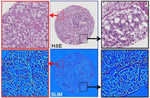
New Way to Look at Tissue Biopsies: Spatial Light Interference Microscopy


Pathologists would gain new tool to diagnose cancer faster and more accurately, based upon stain-free analysis of tissue
Reading tissue biopsies with a new stain-free method could eventually help pathologists achieve faster and less subjective cancer detection. Should this technology prove viable, it would also displace many of the longstanding tissue preparation methodologies used today in the histopathology laboratory.
Credit a research team from the Beckman Institute at the University of Illinois (UI) Christie Clinic and at the UI campuses in Urbana and Chicago, with developing this new technology.
They call the technique Spatial Light Interference Microscopy (SLIM). According to a story reported by Futurity.org, the technique uses two beams of light.
New Technology Could Help Pathologists Detect Cancer Earlier
In the Proceedings of the National Academy of Sciences, the scientists stated the new technology offers answers to some of the most elusive questions in contemporary biology: how cell growth is regulated and how cell size distributions are maintained. “SLIM can be so valuable for greatly improving the chances of early detection and treatment of cancer,” declared study leader Gabriel Popescu, Ph.D., Quantitative Light Imaging Laboratory, Department of Electrical and Computer Engineering at the Beckman Institute.
The reason for Popescu’s optimism is SLIM’s capabilities using optical interferometry, or interference patterns, to make accurate measurements of waves at the molecular level. This enables the technique to work with great sensitivity.

Sunday, January 8, 2012

Pathology Labs Replace Microscopes with Digital Imaging


Non-US deployment of Aperio platform with image storage on a Hitachi platform.  400,000 glass slides annually at a rate of 300 TB of storage per year.  Very cool. Look for more adoption overseas this year.  
Microscopes are being replaced with digital imaging in pathology laboratories in the southern part of Sweden.

Traditional microscope glass slides are turned into digital images, which are then analyzed by pathologists directly from the computer screen, instead of using regular microscopes.

The revolution, which has already occurred in radiology, is now taking place in pathology. The contracted delivery not only digitizes the slides but also will completely renew IT support for all workflows of the pathology laboratories in the Skåne region.

Labvantage (Somerset, NJ, USA) will deliver a USD 4 million turnkey solution for digitizing the histopathological workflows in the whole region. The system will be possibly the largest such installation in the world and among the first of its kind in northern Europe. The digital slides will reside in Hitachi’s (Tokyo, Japan) Content Platform, which employs distributed object storage. All of the images will be kept well protected and duplicated across several physical discs. This makes the traditional backing up of data unnecessary.

Thursday, November 3, 2011

INTERESTING ARTICLES: Damjibhai Anchorwala: Switching to Success

INTERESTING ARTICLES: Damjibhai Anchorwala: Switching to Success: Damjibhai Anchorwala may have dropped his entrepreneurial anchors by selling his Anchor Electricals Company to Panasonic, this Kutchi, in...