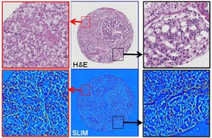
Pathologists would gain new tool to diagnose cancer faster and more accurately, based upon stain-free analysis of tissue
Reading tissue biopsies with a new stain-free method could eventually help pathologists achieve faster and less subjective cancer detection. Should this technology prove viable, it would also displace many of the longstanding tissue preparation methodologies used today in the histopathology laboratory.
Credit a research team from the Beckman Institute at the University of Illinois (UI) Christie Clinic and at the UI campuses in Urbana and Chicago, with developing this new technology.
They call the technique Spatial Light Interference Microscopy (SLIM). According to a story reported by Futurity.org, the technique uses two beams of light.
New Technology Could Help Pathologists Detect Cancer Earlier
In the Proceedings of the National Academy of Sciences, the scientists stated the new technology offers answers to some of the most elusive questions in contemporary biology: how cell growth is regulated and how cell size distributions are maintained. “SLIM can be so valuable for greatly improving the chances of early detection and treatment of cancer,” declared study leader Gabriel Popescu, Ph.D., Quantitative Light Imaging Laboratory, Department of Electrical and Computer Engineering at the Beckman Institute.
The reason for Popescu’s optimism is SLIM’s capabilities using optical interferometry, or interference patterns, to make accurate measurements of waves at the molecular level. This enables the technique to work with great sensitivity.
